Senior Radiologist & Sonologist with 13+ Years of Clinical Precision

")
Clinical Excellence & Diagnostic Precision
A Legacy of Clinical Precision
Specialized Diagnostic Treatments

Whole-Body Health Screening
Proactive wellness checks using advanced ultrasound to detect early-stage changes and establish your healthy baseline.
Book Scan
Routine Ultrasound Imaging
High-precision general imaging for abdominal, pelvic, and small-part assessments to ensure ongoing health monitoring.
Book Scan
Pregnancy Routine Checkup
Essential wellness monitoring and heartbeat checks to ensure a healthy journey for both mother and baby.
Book Scan
NT & Anomaly Screening
Critical developmental assessments, including NT and anomaly scans, performed with extreme detail to ensure fetal well-being.
Book Scan
3D Pelvic Gynaec Ultrasound
Advanced 3D imaging to visualize reproductive organs with exceptional clarity, aiding in the diagnosis of uterine and pelvic conditions.
Book Scan
Pregnancy Scan
A pregnancy scan, or ultrasound, is a safe, non-invasive imaging test using sound waves to create images of the fetus, placenta, and uterus.
Book Scan
Fetal Growth & Doppler Study
Specialized studies tracking growth milestones and vital blood flow dynamics to support a safe delivery plan.
Book Scan
3D & 4D Obstetric Vision
Experience a life-like connection with your baby through high-definition 3D/4D visualization of fetal anatomy.
Book Scan
Expert Sonomammography
Advanced breast ultrasound and mammography evaluations for precise identification of clinical diagnostic concerns.
Book Scan
Mass Localization & Clip Insertion
Guided identification and clip localization of small breast masses for precise duration-based clinical mapping.
Book Scan
Specialized Onco-Imaging
Targeted oncological screening protocols focused on early detection, staging, and diagnostic monitoring of concerns.
Book Scan
Guided Fluid Aspiration
Minimally invasive, ultrasound-guided drainage of pleural, ascitic, or other fluid collections for analysis.
Book Scan
USG-Guided FNAC
High-precision Fine Needle Aspiration Cytology for accurate tissue diagnosis with minimal local intervention.
Book Scan
PCN For Renal Abscess
High-precision Fine Needle Aspiration Cytology for accurate tissue diagnosis with minimal local intervention.
Book Scan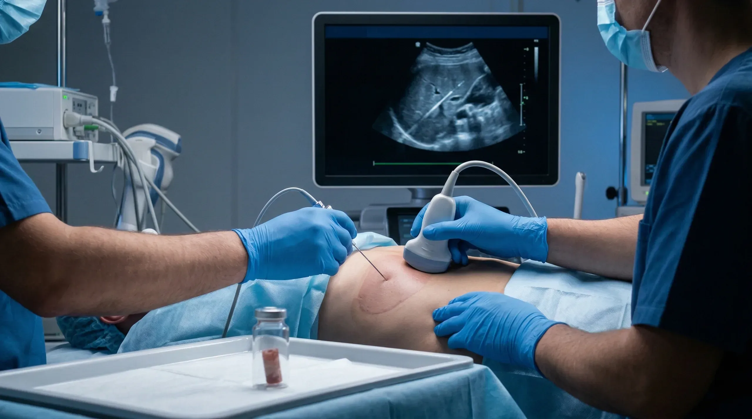
Multi-Organ Guided Biopsy
Advanced guided tissue sampling for Liver, Kidney, Breast, and Lymph Nodes using real-time visualization.
Book Scan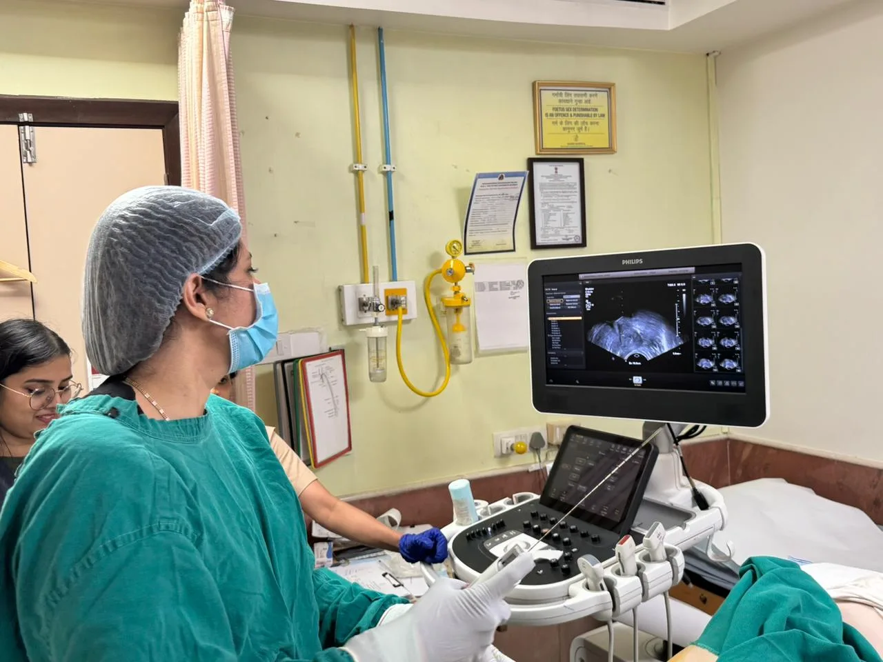
TRUS Guided Prostate Biopsy
Specialized transrectal ultrasound-guided sampling for definitive diagnostic accuracy in prostate care.
Book Scan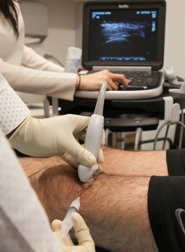
Guided Steroid Injections
Targeted pain relief for chronic muscular conditions via precision-guided intramuscular injections.
Book Scan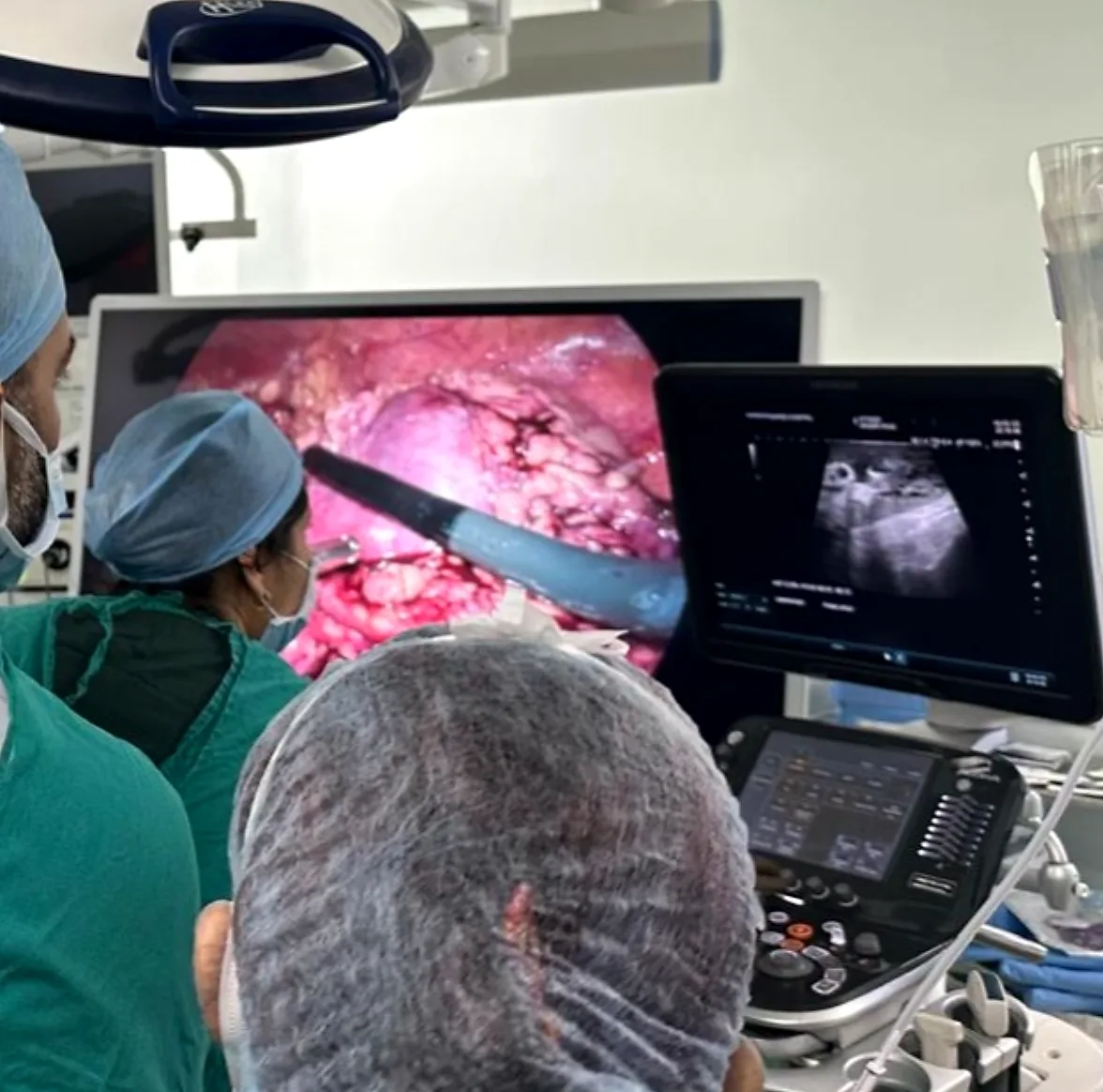
Intraoperative Ultrasound
Real-time intra-operative imaging providing high-precision visual data for complex surgical interventions.
Book Scan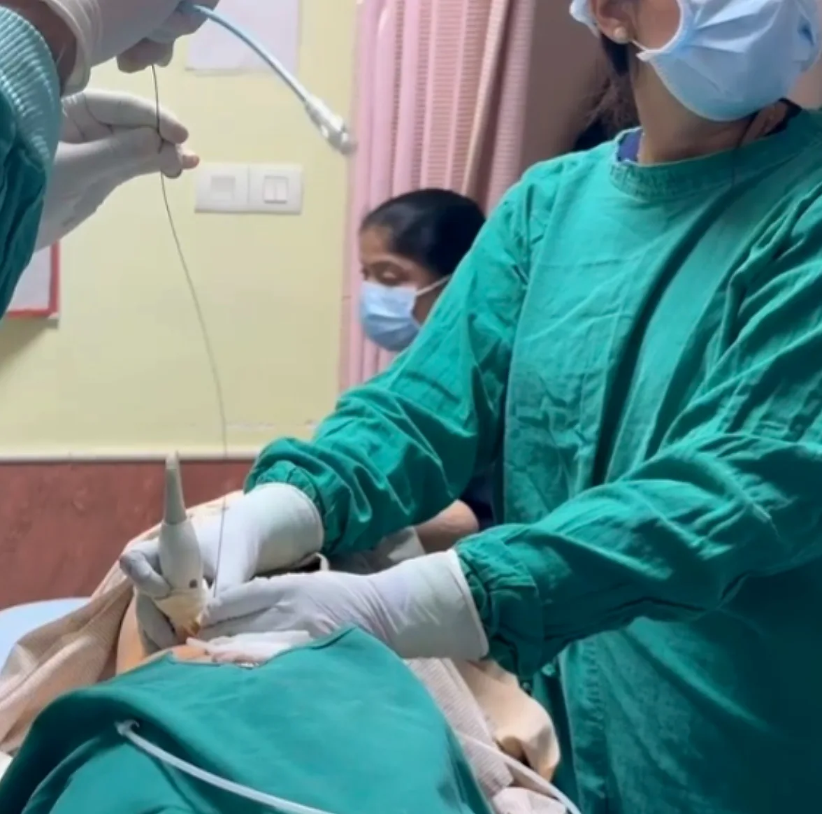
Pigtail Catheterization
Percutaneous drainage of abscesses and fluid collections using advanced imaging guidance.
Book Scan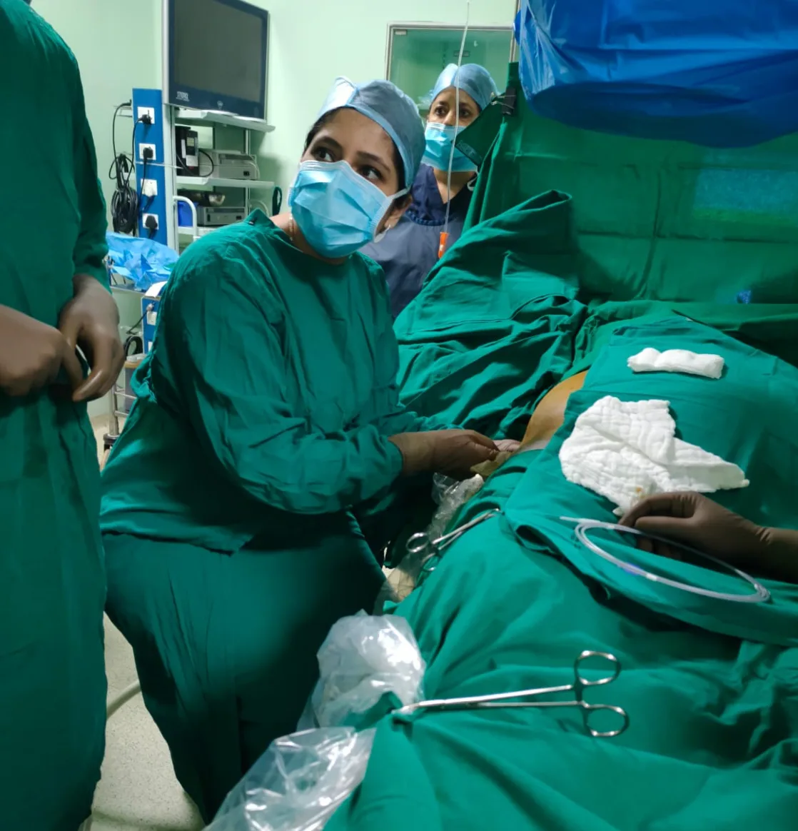
(IOUS) Tumor Localization
Intraoperative ultrasound (IOUS) tumor localization is a real-time, cost-effective imaging technique used during surgery to precisely locate tumors.
Book ScanWhat Makes Dr. Shehzia Lakhani Different


Answers that help you
decide with confidence
Get in touch with us
You can connect with us now by clicking here: Connect via WhatsApp.
Alternatively, reach us via email at hello@drshehzialakhani.com or visit us at Allbless Diagnostic Centre, Mumbai. Early booking is recommended for specialized scans.
Ultrasound (USG) is a very important screening tool during pregnancy and can detect many structural abnormalities in the fetus. However, not all anomalies can be identified through ultrasound alone.
Some conditions may be too small to detect or may develop later in pregnancy. Regular scans performed by an experienced radiologist like Dr. Shehzia Lakhani help maximize the chances of early detection and monitoring.
Color Doppler is a specialized ultrasound technique used to visualize and measure blood flow in vessels. It helps doctors evaluate circulation in arteries and veins and is commonly used in pregnancy, heart studies, and vascular conditions. In pregnancy, it can assess blood flow between the mother, placenta, and baby.
Many early breast changes cannot be felt during a physical examination. A mammogram can detect very small abnormalities in breast tissue years before they become noticeable lumps. This makes mammography an important screening tool for early detection of breast cancer, when treatment is most effective.
Regular health checkups allow doctors to identify potential health problems early, often before symptoms appear. Early detection helps in better treatment outcomes and prevents complications. Imaging tests like ultrasound and mammography are often part of routine preventive healthcare.
Yes, ultrasound is considered very safe during pregnancy. It uses sound waves rather than radiation, making it a safe imaging method for both the mother and the baby when performed by trained professionals.
The number of ultrasounds during pregnancy depends on medical needs and your doctor’s recommendation. In most pregnancies, a few key scans are recommended at specific stages, but additional scans may be advised if closer monitoring is required.
Some important scans during pregnancy include:
- Early Pregnancy Scan – Confirms pregnancy location, heartbeat, and gestational age.
- NT Scan (First Trimester Scan) – Screens for certain chromosomal abnormalities.
- Anomaly Scan (18–22 weeks) – Examines the baby’s organs and structure to detect possible abnormalities.
- Growth Scan – Monitors the baby’s growth, position, and amniotic fluid levels later in pregnancy.
- Obstetric Doppler Scan – Routinely performed during pregnancy to evaluate blood flow between the mother, placenta, and baby, helping ensure the baby is receiving adequate oxygen and nutrients.
Each scan plays a crucial role in ensuring the health and development of the baby.
Ureteric stones can occasionally be difficult to detect on ultrasound because the ureters are thin tubes and may be obscured by bowel gas or body structures. In some cases, additional imaging such as a CT scan may be recommended for a clearer diagnosis.
A transvaginal ultrasound involves placing a small ultrasound probe inside the vagina to obtain clearer images of the uterus, ovaries, and early pregnancy structures. It is especially helpful for early pregnancy evaluation, fertility assessments, and detailed pelvic imaging.
No, the majority of cysts are benign (non-cancerous). Many cysts are common and harmless, especially ovarian cysts or simple cysts seen on imaging. However, imaging tests and medical evaluation are important to determine the nature of the cyst and whether any follow-up is needed.
Dr. Shehzia Lakhani is an experienced radiologist specializing in diagnostic imaging, ultrasound, pregnancy scans, and breast imaging. She focuses on providing accurate reports and clear guidance to support both patients and referring doctors.
Dr. Shehzia Lakhani offers a range of diagnostic imaging Treatments including:
- Pregnancy ultrasounds
- Anomaly scans
- Color Doppler studies
- Breast imaging and mammography
- General ultrasound examinations
- Ultrasound guided interventions
Patients choose Dr. Shehzia Lakhani for her experience, diagnostic accuracy, patient-centered approach, and detailed reporting. Her focus is on providing reliable imaging interpretations to support better medical decisions.
Yes, for most diagnostic scans, a valid doctor's prescription is required as per medical regulatory guidelines. Specifically for pregnancy scans, a filled Form F is mandatory under the PC-PNDT Act.
Preparation depends on the type of scan. For Upper Abdomen scans, 6-8 hours of fasting is required. For Pelvic or Early Pregnancy scans, you need a full bladder. Our team will provide precise instructions at the time of booking.
We value your time. Most ultrasound reports are prepared and handed over within 30-60 minutes of the procedure, ensuring you can proceed with your medical consultation without unnecessary delay.
Get in touch with us
